
Lameness

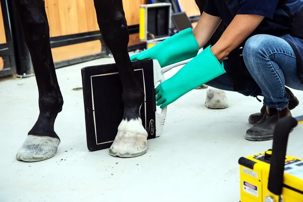
Lameness diagnostics and therapeutics are rapidly advancing areas in equine sports medicine. Lameness is the most common cause of limiting performance, and can range from being very obvious to extremely subtle. We utilize multiple modalities including palpation, joint and nerve anesthesia, digital radiography and digital ultrasonography to locate the cause of the lameness. We then work with you, based on your goals and expectations, to customize the appropriate therapeutic and rehabilitative plan for your horse.
Diagnostics
Swiftsure Equine Veterinary Services offers diagnostic imaging to help identify the source of lameness and guide effective treatment planning. Our diagnostic tools include digital radiography (X-ray) and ultrasonography, allowing us to evaluate bones, joints, soft tissues, tendons, and ligaments with precision.
These imaging modalities help us accurately diagnose many common causes of lameness and develop targeted treatment and rehabilitation plans to support your horse’s recovery and performance.
-
X-rays (radiographs) are the most common tools our veterinarians use to evaluate bones and joints in horses. They work by passing a small amount of radiation through the body to create images of dense bone structure.
What X-rays are used for:
Identifying fractures or bone chips
Evaluating joint health
Detecting signs of arthritis
Assessing hoof balance and structures (like the coffin bone)
Investigating causes of lameness
How they detect injury:
X-rays show clear images of bone, so vets can spot:
Breaks or cracks in bone
Bone fragments or chips within a joint
Abnormal bone growth or remodeling
Changes caused by repeated stress or trauma
How they detect arthritis:
In horses, arthritis (degenerative joint disease) causes visible changes over time. X-rays can reveal:
Narrowing of joint spaces (loss of cartilage)
Bone spurs (new bone growth around joints)
Increased bone density or irregular joint surfaces
Chronic changes that indicate long-term wear and tear
Limitations:
X-rays do not show soft tissues like tendons and ligaments very well, so they’re often used alongside ultrasound or other imaging methods for a complete picture.
Overall, X-rays are a fast, effective way to diagnose bone injuries and monitor joint conditions like arthritis, helping veterinarians develop the right treatment and management plan for your horse.
-
Ultrasound is a non-invasive imaging tool used in horses to look at soft tissues beneath the skin and detect injuries that can’t be appreciated from other modalities such as radiographic imaging.
During the procedure, a veterinarian places a handheld probe (transducer) on the horse’s skin—usually over the area of concern. The probe sends sound waves into the tissue, and those waves bounce back to create real-time images on a screen.
Ultrasound is especially useful for examining:
Tendons and ligaments (like suspensory or flexor tendon injuries)
Muscles and soft tissue damage
Joint structures and fluid buildup
Some areas of the reproductive tract or internal organs
It helps detect injuries by showing:
Tears or lesions in fibers
Swelling or inflammation
Changes in tissue structure or alignment
Fluid accumulation
Ultrasound is also commonly used to monitor healing over time, allowing veterinarians to adjust rehabilitation plans based on how the tissue is repairing.
Overall, it’s a safe, painless, and highly effective way to diagnose and track soft tissue injuries in horses.
Treatments
Once the source of lameness has been identified, we develop a targeted treatment plan based on your horse’s diagnosis, intended use, and long-term goals.
Treatment options may include a combination of medical therapies, regenerative medicine, and supportive care, all designed to reduce pain, promote healing, and support a safe return to work or performance.
-
Joint injections are used to reduce inflammation and manage pain within affected joints by delivering medication directly to the source of discomfort. This targeted approach can help improve mobility, maintain performance, and support joint health.
They are commonly recommended for horses experiencing joint inflammation, arthritis, or performance-related wear, and are selected based on each horse’s specific needs and level of activity.
-
Shockwave therapy (often called extracorporeal shockwave therapy, or ESWT) is a non-invasive treatment used in horses to help heal certain injuries.
Shockwave delivers controlled sound waves into injured tissue. These waves stimulate the body’s natural healing processes by increasing blood flow, reducing inflammation, and encouraging the repair of damaged cells. It can also provide pain relief by temporarily desensitizing nerve endings in the treated area.
Shockwave is used for conditions involving tendons, ligaments, and joints, wounds, and bone stress injuries. Common locations include strained tendons or ligaments, ring bone, popped splints, sore necks and backs, and wounds. Overall, it helps speed up recovery, improve comfort, and support better healing without surgery, though it’s usually combined with rest and a proper rehabilitation plan.
-
PRP, or Platelet Rich Plasma is a concentrate of the platelets from a sample of the horse’s own blood, taken sterilely and processed in a specific manner. Platelets are a very powerful component of blood which, when activated, release a variety of growth factors and cytokines to attract other healing cells to the area. If a tendon or a ligament has a “core lesion” within it, it typically is an excellent candidate for PRP therapy. Injecting PRP into a tendon or ligament injury can both speed the healing process and help the injury heal more effectively (ie with less scar tissue), making it less prone to reinjury in the future. The lesion in the tendon/ligament is injected under ultrasound guidance so the veterinarian can be sure the PRP is being injected into exactly the right spot within the injury. The PRP treatment is best used in combination with a tailored rest and rehabilitation program to give the horse the very best chance at a full recovery.
-
ProStride, is an autologous protein solution, which effectively combines PRP with naturally occurring anti-inflammatory molecules like Interleukin-1 Receptor Antagonist Protein (IRAP) and Tumor Necrosis Factor (TNF-α). This makes for a potent anti-inflammatory product that also promotes repair of damaged tissues. Like the PRP, this product is made from a sample of the horse’s own blood, processed in a specific way. ProStride makes an excellent alternative to steroid joint injections for those horses who cannot safely have steroids due to metabolic disease, or who have a combination of arthritis and soft tissue injury, or who are not responding to intra-articular steroids as well as they once were.
-
Centrate BMA is a bone marrow aspiration device designed to concentrate regenerative cells from the horse's own bone marrow. It is an advanced regenerative therapy used in horses to support healing in joints and surrounding structures.
In this procedure, bone marrow— taken from the horse’s sternum — is collected and then processed (centrifuged) to concentrate its most beneficial components, including stem cells, growth factors, and anti-inflammatory proteins. This concentrated material is then injected directly into the affected joint(s).
These injections are used to:
Reduce inflammation within the joint
Promote repair of damaged cartilage and soft tissue
Support healing in more serious or chronic injuries
Potentially slow the progression of joint conditions like arthritis
Because bone marrow contains stem cells, this therapy goes a step further than standard blood-based treatments by actively supporting tissue regeneration, not just reducing inflammation.
It’s commonly used for horses with more significant joint damage, chronic lameness, or injuries that haven’t responded well to other treatments. As with other regenerative therapies, it’s typically combined with a structured rehabilitation program for the best results.
Resources for Clients